Director's Message - Spring 2023

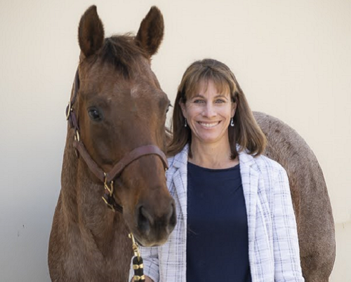
I hope that the longer days and sunnier spring weather have been translating into more time with your horses. I am personally enjoying a return to three-day eventing competitions after a very wet winter here in Northern California.
The focus of this issue of the Horse Report, diagnostic imaging, is important to me professionally as a veterinarian and researcher and personally as a horse owner. It has been more than a year since my Thoroughbred gelding, Chat, sustained a coffin bone fracture. The diagnosis and treatment plans for his injury relied on X-rays, which were performed at the UC Davis veterinary hospital. I am happy to say that Chat is now in full work after repeated follow-up exams with our imaging team. We are incredibly fortunate to have advanced imaging equipment and highly trained clinicians and veterinary technicians on hand to provide this important service at UC Davis.
Our faculty contributors for this issue - Drs. Kathryn Phillips, Mathieu Spriet, Betsy Vaughan and Mary Beth Whitcomb - are experts in all forms of equine diagnostic imaging. In addition to providing top-of-the-line care for patients, they also conduct research to advance imaging approaches and applications, which continues to make UC Davis a leader in this area. We are grateful for their time and expertise to provide the most up-to-date information on this topic.
From X-rays to CT, our goal for this issue is to demystify these commonly performed but often poorly understood technologies. Diagnostic imaging is central to diagnosis and treatment in many cases. Understanding the basics of the different modalities can help you more effectively work with your veterinarian to determine the best approaches for your horses when they are ill or injured.
Best wishes,

Thanks To Our Collaborators

Kathryn Phillips, DVM, DACVR, DACVR-EDI – Dr. Phillips is a double board-certified radiologist with expertise in diagnostic imaging in a variety of species. Her research focus is on equine and exotic animal imaging.

Mathieu Spriet, DVM, MS, DACVR, DECVDI, DACVR-EDI – Dr. Spriet is a multiple board-certified radiologist. His research focus is in advanced musculoskeletal imaging in horses, particularly the development of PET imaging for lameness assessment in sport horses and prevention of catastrophic breakdown in racehorses.

Betsy Vaughan, DVM, DACVSMR – An expert in large animal ultrasound, Dr. Vaughan’s interests include equine musculoskeletal injuries and rehabilitation, as well as equine and livestock abdominal imaging.

Mary Beth Whitcomb, DVM, MBA, ECVDI(LA-Associate) – Professor Emeritus Whitcomb is an expert in large animal ultrasound. Her research focus is ultrasonographic techniques and educational models for the diagnosis of upper limb lameness in horses.
