UC Davis Imaging Services
Diagnostic imaging services for horses at the UC Davis veterinary hospital include radiography (X-ray), ultrasound, computed tomography (CT), magnetic resonance imaging (MRI), nuclear scintigraphy (“bone scan”), and positron emission tomography (PET). Imaging is fundamental to patient care across multiple specialties, including surgery, neurology, and ophthalmology.
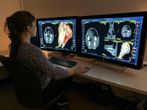
UC Davis is at the forefront of advances in diagnostic imaging technology. It is one of only a few veterinary hospitals in the country with a service dedicated exclusively to large animal ultrasound and staffed by faculty specifically Dr. Charlene Pige analyzing a CT scan of a horse’s head. trained in its use. Advanced imaging capabilities for horses include both standing PET and MRI. The planned acquisition of a new CT unit with the ability to image the equine head and neck will provide additional diagnostic options for patients.
In the future, the All Species Imaging Center, currently under construction as part of the new Veterinary Medical Center, will act as a central hub of imaging services for all hospital specialties. Diagnostic imaging specialists, accompanied by the largest team of residents at any veterinary hospital, will utilize the most cutting-edge elements of imaging techniques such as CT, MRI, nuclear scintigraphy, and PET scanning. Advanced technologies and clinical research will provide innovative detection, diagnosis, and treatment of disease and trauma.